
Life Sciences
| Liquid Flow TEM | Liquid Electrochemistry TEM | Liquid Heating TEM | Liquid X-Ray | Liquid SEM | ||
| Imaging | Live imaging |  |
 |
 |
 |
 |
| Higher resolution |  |
 |
 |
 |
 |
|
| EDS/EELS compatibility |  |
 |
 |
 |
 |
|
| 3D reconstruction |  |
 |
 |
 |
 |
|
| Cross-correlative microscopy |  |
 |
 |
 |
 |
|
| Thermo – Response | Thermal cycling |  |
 |
 |
 |
 |
| Reliable temperature input/output |  |
 |
 |
 |
 |
|
| Quantitative Analysis | Particle statistics |  |
 |
 |
 |
 |
| Biasing input/output |  |
 |
 |
 |
 |
|
 Excellent
  Good
Good  N/A
N/A
Electron holography of bacterial cell in liquid
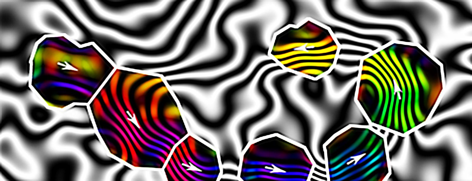
Researchers at U.S. Ames Laboratory, Imperial College London and Ernst Ruska-Center for Microscopy and Spectroscopy with Electrons and Peter Grunberg Institute have used Hummingbird Scientific’s continuous liquid cell platform to demonstrate the first holographic imaging of bacterial and magnetic particles in liquid.
The effect of electromagnetic fields in the biological system has been poorly understood. The researcher performed off-axis holography imaging of hydrated cells of Magentospirillum magneticum strain AMB-1 and assemblies of magnetic nanoparticles. They were able to capture electron holograms to show interference fringe contrast to allow reconstruction of the phase shift of the electron wave and mapping of the magnetic induction from bacterial magnetite nanocrystals. The development of this technique could potentially help in the future studies of solid-liquid interfaces, biomineralization and protein aggregation.
Reference: Tanya Prozorov et al. Off-axis electron holography of bacterial cells and magnetic nanoparticles in liquid. Journal of The Royal Society Interface (2017). Abstract
Top page banner: Image copyright © 2017 The Royal Society Publishing
Edit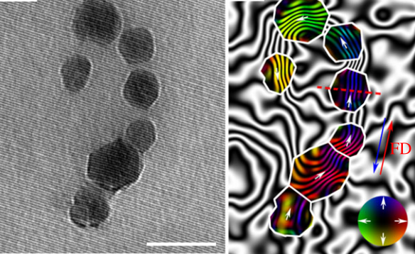
Off-axis electron hologram and magnetic induction map of nanocrystal chain of ruptured bacterium. The scale bar in 100 nm. Image copyright © 2017 The Royal Society Publishing
Growing and imaging micelle assembly
The mechanism of micelle growth and assembly has been a subject of much investigations. There are several theories explaining the assembly process of micelle in liquid such as unimer insertion, fusion, fission, complete micelle disassembly and reassembly, or nucleation-elongation. The researchers led by Northwestern University used in-situ liquid cell transmission electron microscopy to monitor the motion and evolution of individual micelles in real time with nanometer spatial resolution. This liquid platform allowed them to observe the diffusion behavior, fusion events and micelle-micelle interaction in solution. This direct observation of micelle interaction in solution opens up a new avenue to test the evolution of soft matter assemblies in unconstrained liquid medium.
Video shows micelle-micelle fusion event in liquid cell TEM.
Reference: Nathan C. Gianneschi et al. Directly Observing Micelle Fusion and Growth in Solution by Liquid-Cell Transmission Electron Microscopy. Journal of the American Chemical Society (2018). Abstract
Movie copyright © 2017 American Chemical Society
Edit| Feng Yan, Lili Liu, Tiffany R. Walsh, Yu Gong, Patrick Z. El-Khoury, Yanyan Zhang, Zihua Zhu, James J. De Yoreo, Mark H. Engelhard, Xin Zhang, and Chun-Long Chen . “Controlled synthesis of highly-branched plasmonic gold nanoparticles through peptoid engineering,” Nature Communications (2018) | Abstract |
| Peter Sutter, Bo Zhang, and Eli Sutter. “Radiation damage during in-situ electron microscopy of DNA-mediated nanoparticle assemblies in solution.” Nanoscale (2018) | Abstract |
| Alejandra Londono-Calderon, Lee Bendickson, Pierre E. Palo, Marit Nilsen-Hamilton, Surya Mallapragada and Tanya Prozorov. “In-Situ Nucleation, Growth and Evolution of Au Nanoparticles during Metallization of DNA Origami Visualized with HAADF-STEM.” Microscopy & Microanalysis (2018) | Abstract |
| Lucas R. Parent, Evangelos Bakalis, Abelardo Ramírez-Hernández, Jacquelin K. Kammeyer, Chiwoo Park, Juan de Pablo, Francesco Zerbetto, Joseph P. Patterson, and Nathan C. Gianneschi.”Directly Observing Micelle Fusion and Growth in Solution by Liquid-Cell Transmission Electron Microscopy,” Journal of the American Chemical Society (2017) | Abstract |
| Tanya Prozorov, Trevor P. Almeida, András Kovács, and Rafal E. Dunin-Borkowski. ” Off-axis electron holography of bacterial cells and magnetic nanoparticles in liquid,” Journal of The Royal Society Interface (2017) | Abstract |
| Arjan P. H. Gelissen, Alex Oppermann, Tobias Caumanns, Pascal Hebbeker, Sarah K. Turnhoff, Rahul Tiwari, Sabine Eisold, Ulrich Simon, Yan Lu, Joachim Mayer, Walter Richtering, Andreas Walther, and Dominik Wöll.”3D Structures of Responsive Nanocompartmentalized Microgels,” Nano Letters (2016) | Abstract |
| J. P. Patterson, L. R. Parent, J. Cantlona, H. Eickhoffa, G. Bareda, J. E. Evansa and N.C. Gianneschia. “Picoliter Drop-On-Demand Dispensing for Multiplex Liquid Cell Transmission Electron Microscopy,” Microscopy and Microanalysis, Online (2016) | Abstract |
| T.J. Woehl, S. Kashyap, E. Firlar, T. Perez-Gonzalez, D. Faivre, D. Trubitsyn, D. Bazylinski, T. Prozorov. “Correlative Electron and Fluorescence Microscopy of Magnetotactic Bacteria in Liquid: Toward In-Vivo Imaging,” Microscopy and Microanalysis (2015) | Abstract |
| P. J. M. Smeets, K. R. Cho, R. G. E. Kempen, N. A. J. M. Sommerdijk, and J. J. De Yoreo. “Calcium carbonate nucleation driven by ion binding in a biomimetic matrix revealed by in situ electron microscopy”, Nature Materials Letters, Published Online 01/26/2015. | Abstract |
| M.H. Nielsen, S. Aloni, J.J. De Yoreo. “In situ TEM imaging of CaCO3 nucleation reveals coexistence of direct and indirect pathways”, Science vol. 345 iss. 6201 (2014) pp. 1158-1162 | Abstract |
| S. Kashyap, T.J. Woehl, X. Liu, S.K. Mallapragada, T. Prozorov. ”Nucleation of Iron Oxide Nanoparticles Mediated by Mms6 Protein In Situ“ ACS Nano (2014) | Abstract |
| D.A. Fischer, D.H. Alsem, B. Simon, T. Prozorov and N. Salmon. “Development of an Integrated Platform for Cross-Correlative Imaging of Biological Specimens in Liquid using Light and Electron Microscopies.” Microscopy and Microanalysis (2013) | Abstract |
| F.A. Plamper, A.P. Gelissen, J. Timper, A. Wolf, A.B. Zezin, W. Richtering, H. Tenhu, U. Simon, J. Mayer, O. V. Borisov, D.V. Pergushov. “Spontaneous Assembly of Miktoarm Stars into Vesicular Interpolyelectrolyte Complexes,” Macromolecular Rapid Communication (2013) | Abstract |
| J.E. Evans, K.L. Jungjohann, P.C.K. Wong, P.L. Chiua, G.H. Dutrowa, I. Arslan, N.D. Browning. “Visualizing macromolecular complexes with in-situ liquid scanning transmission electron microscopy” Micron (2012) | Abstract |
| J.E. Evans, N.D. Browning, “Enabling Direct Nanoscale Observations of Biological Reactions with Dynamic TEM,” Microscopy (2013) | Abstract |
| N. de Jonge, D.B. Peckys, G.J. Kremers, D.W. Piston. “Electron microscopy of whole cells in liquid with nanometer resolution,” PNAS (2009) | Abstract |
Read More


